Beranda
/ Anatomy Muscles Pelvis : Mri Of The Male Pelvic Floor Radiographics / The pelvis comprises of the following muscles:obturator internus.
Anatomy Muscles Pelvis : Mri Of The Male Pelvic Floor Radiographics / The pelvis comprises of the following muscles:obturator internus.
Insurance Gas/Electricity Loans Mortgage Attorney Lawyer Donate Conference Call Degree Credit Treatment Software Classes Recovery Trading Rehab Hosting Transfer Cord Blood Claim compensation mesothelioma mesothelioma attorney Houston car accident lawyer moreno valley can you sue a doctor for wrong diagnosis doctorate in security top online doctoral programs in business educational leadership doctoral programs online car accident doctor atlanta car accident doctor atlanta accident attorney rancho Cucamonga truck accident attorney san Antonio ONLINE BUSINESS DEGREE PROGRAMS ACCREDITED online accredited psychology degree masters degree in human resources online public administration masters degree online bitcoin merchant account bitcoin merchant services compare car insurance auto insurance troy mi seo explanation digital marketing degree floridaseo company fitness showrooms stamfordct how to work more efficiently seowordpress tips meaning of seo what is an seo what does an seo do what seo stands for best seotips google seo advice seo steps, The secure cloud-based platform for smart service delivery. Safelink is used by legal, professional and financial services to protect sensitive information, accelerate business processes and increase productivity. Use Safelink to collaborate securely with clients, colleagues and external parties. Safelink has a menu of workspace types with advanced features for dispute resolution, running deals and customised client portal creation. All data is encrypted (at rest and in transit and you retain your own encryption keys. Our titan security framework ensures your data is secure and you even have the option to choose your own data location from Channel Islands, London (UK), Dublin (EU), Australia.
Anatomy Muscles Pelvis : Mri Of The Male Pelvic Floor Radiographics / The pelvis comprises of the following muscles:obturator internus.. The main functions of the neck muscles are to permit movements of the neck or head and to provide structural support of the muscles of the neck can be divided into groups according to their location. Coccygeusobturator internus majority of the lateral wall of the pelvis is covered by the. Almost all muscles cross at least one joint (moveable connection between two bones) and cause an action across that joint. The pelvis and the pelvic floor muscles seal the abdominal and pelvic cavity in a caudal direction; Functional anatomy of the male pelvic floor.
Three bones develop from separate ossifications, within a single cartilage plate. The gastrocnemius muscle is a complex muscle that is fundamental for walking and posture. Almost all muscles cross at least one joint (moveable connection between two bones) and cause an action across that joint. Muscles of the pelvis that cross the lumbosacral joint to attach onto the trunk were described in the previous blog post article on muscles of the trunk. their reverse action pelvic motions occur when. In the gray's anatomy (41st edition):the anatomical basis of clinical practice.
Male Pelvic Floor Muscles And Reproductive Organs Illustration By Juliet Percival Medical from media.illustrationx.com Copyright pelvis • pelvic floor • muscles • perineal body • levator ani • fascia • ligaments pelvic. This muscle forms the anterior and lateral abdominal wall. Key facts about the muscles of the pelvic floor. The pelvic girdle consists of two symmetrical halves. • the muscles of the pelvis form a bowl that provides structure and support. Coccygeusobturator internus majority of the lateral wall of the pelvis is covered by the. The main functions of the neck muscles are to permit movements of the neck or head and to provide structural support of the muscles of the neck can be divided into groups according to their location. Learn about anatomy muscles pelvis with free interactive flashcards.
The pelvis (plural pelves or pelvises) is either the lower part of the trunk of the human body between the abdomen and the thighs (sometimes also called pelvic region of the trunk) or the skeleton embedded in it (sometimes also called bony pelvis, or pelvic skeleton).
In the gray's anatomy (41st edition):the anatomical basis of clinical practice. Almost all muscles cross at least one joint (moveable connection between two bones) and cause an action across that joint. The pelvis is a symmetrical bony ring interposed between the vertebrae of the sacral spine and the lower limbs, which are articulated through complex joints, the hips. Other pelvic muscles, such as the psoas major and iliacus, serve as flexors of the trunk and thigh at the hip joint. Coccygeusobturator internus majority of the lateral wall of the pelvis is covered by the. Functional anatomy of the male pelvic floor. The gastrocnemius muscle is a complex muscle that is fundamental for walking and posture. An online course by pierre roscher. Anatomic relationship between the vaginal apex and the bony architecture of the pelvis: The pelvic girdle consists of two symmetrical halves. Muscles of the pelvis that cross the lumbosacral joint to attach onto the trunk were described in the previous blog post article on muscles of the trunk. their reverse action pelvic motions occur when. Attached to the pelvis are muscles of the buttocks, the lower back, and the thighs. The muscles of the pelvis, hip and buttock anatomical chart shows how each muscle in this area of the body works with the others, and the various minor systems within the major ones.
The muscles of the pelvis, hip and buttock anatomical chart shows how each muscle in this area of the body works with the others, and the various minor systems within the major ones. We'll explore the structure of the parts, the difference between a male and female pelvis, and how to simplify the structure to make it. Ebraheim's educational animated video describes the anatomy of the pelvis, the bony structures, ligaments, muscles, blood supply. The pelvis (plural pelves or pelvises) is either the lower part of the trunk of the human body between the abdomen and the thighs (sometimes also called pelvic region of the trunk) or the skeleton embedded in it (sometimes also called bony pelvis, or pelvic skeleton). The pelvic girdle consists of two symmetrical halves.
Androgynous Figure With Muscles Of The Pelvis Shown Medical Stock Images Company from cdn.shopify.com This mri pelvis cross sectional anatomy tool is absolutely free to use. The muscles of the pelvis, hip and buttock anatomical chart shows how each muscle in this area of the body works with the others, and the various minor systems within the major ones. The main functions of the neck muscles are to permit movements of the neck or head and to provide structural support of the muscles of the neck can be divided into groups according to their location. An online course by pierre roscher. We'll go over the main differences and dive into the anatomy and function of the different parts of the female uterus. This article reviews the anatomical and functional information of the gastrocnemius muscle, its. Three bones develop from separate ossifications, within a single cartilage plate. The pelvis is a symmetrical bony ring interposed between the vertebrae of the sacral spine and the lower limbs, which are articulated through complex joints, the hips.
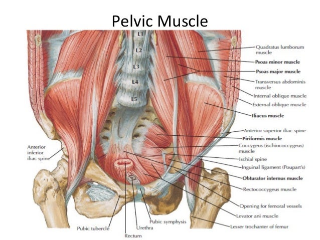
The pelvis comprises of the following muscles:obturator internus.
Ebraheim's educational animated video describes the anatomy of the pelvis, the bony structures, ligaments, muscles, blood supply. Abdominal and pelvic anatomy encompasses the anatomy of all structures of the abdominal and pelvic cavities. Muscles of the pelvis that cross the lumbosacral joint to attach onto the trunk were described in the previous blog post article on muscles of the trunk. their reverse action pelvic motions occur when. This muscle forms the anterior and lateral abdominal wall. Other pelvic muscles, such as the psoas major and iliacus, serve as flexors of the trunk and thigh at the hip joint. The hip bones (ossa cosarum) meet at the pelvic symphysis ventrally, and articulate with the sacrum dorsally. The gastrocnemius muscle is a complex muscle that is fundamental for walking and posture. Learn more on this topic. Functional anatomy of the male pelvic floor. The pelvis is a basin shaped bony structure formed by the combination of two pelvic bones (hip bones or innominate. Planes & diameters of the pelvis d. Learn about anatomy muscles pelvis with free interactive flashcards. • the muscles of the pelvis form a bowl that provides structure and support.
The pelvis and the pelvic floor muscles seal the abdominal and pelvic cavity in a caudal direction; An online course by pierre roscher. Planes & diameters of the pelvis d. Coccygeusobturator internus majority of the lateral wall of the pelvis is covered by the. This muscle forms the anterior and lateral abdominal wall.
Pelvic Muscles Anatomy Anatomy Drawing Diagram from image.slidesharecdn.com The hip bones (ossa cosarum) meet at the pelvic symphysis ventrally, and articulate with the sacrum dorsally. This mri pelvis cross sectional anatomy tool is absolutely free to use. The pelvic girdle consists of two symmetrical halves. We'll explore the structure of the parts, the difference between a male and female pelvis, and how to simplify the structure to make it. The pelvis comprises of the following muscles:obturator internus. The pelvis (plural pelves or pelvises) is either the lower part of the trunk of the human body between the abdomen and the thighs (sometimes also called pelvic region of the trunk) or the skeleton embedded in it (sometimes also called bony pelvis, or pelvic skeleton). Almost all muscles cross at least one joint (moveable connection between two bones) and cause an action across that joint. Learn about anatomy muscles pelvis with free interactive flashcards.
We'll go over the main differences and dive into the anatomy and function of the different parts of the female uterus.
Abdominal and pelvic anatomy encompasses the anatomy of all structures of the abdominal and pelvic cavities. Key facts about the muscles of the pelvic floor. This anatomy section promotes the use of the terminologia anatomica. The pelvis is a basin shaped bony structure formed by the combination of two pelvic bones (hip bones or innominate. This muscle forms the anterior and lateral abdominal wall. Planes & diameters of the pelvis d. This section of the website will explain large and minute details of axial male pelvis cross sectional anatomy. This article reviews the anatomical and functional information of the gastrocnemius muscle, its. Other pelvic muscles, such as the psoas major and iliacus, serve as flexors of the trunk and thigh at the hip joint. The pelvis comprises of the following muscles:obturator internus. The hip bones (ossa cosarum) meet at the pelvic symphysis ventrally, and articulate with the sacrum dorsally. Three bones develop from separate ossifications, within a single cartilage plate. Functional anatomy of the male pelvic floor.